
Caring for breast health requires ongoing attention and medical follow-up throughout life. Radiologist Vinicius Tadeu Sattin Rodrigues explains that mammography allows for a highly detailed analysis of breast tissue, helping identify structural characteristics that would not be detected through clinical examination alone. For this reason, the exam has taken on an important role in women’s preventive health strategies.
Although it is widely known, many people still do not fully understand how mammography contributes to preventive monitoring. Gaining a better understanding of how this method works helps in interpreting results and medical recommendations related to breast health. In this article, we will explore how mammography assists in observing the breasts and why it is considered essential for preventive monitoring. Read this article to the end to learn more about the topic.
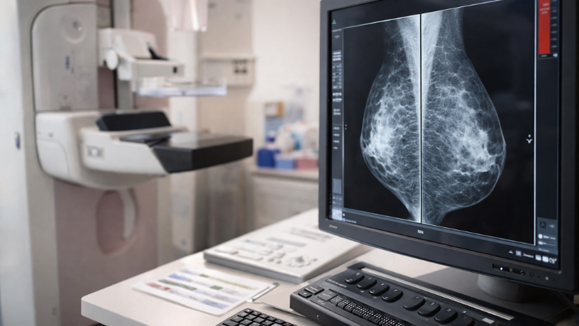
How does mammography allow the observation of breast structure?
Mammography uses radiological technology capable of generating detailed images of the inside of the breasts. Former Health Secretary Vinicius Tadeu Sattin Rodrigues states that the exam makes it possible to visualize different types of tissue present in the breast, such as glandular, fibrous, and adipose structures. These structures appear in the images with distinct characteristics, making analysis easier for the radiologist.
During image interpretation, the specialist observes structural patterns and possible formations within the breast tissue. This evaluation helps to better understand breast composition. In this context, mammography becomes an important tool for analyzing the interior of the breasts. The detailed observation of the images contributes to a more comprehensive assessment of breast health.

What structures can appear in mammography images?
Images obtained during mammography may reveal different characteristics of breast tissue. Among the elements observed are small calcifications, nodules, and variations in breast density. According to Vinicius Tadeu Sattin Rodrigues, each of these structures is carefully analyzed to understand its nature.
Comparing mammograms taken at different times is part of radiological analysis. This process allows for the observation of possible changes in breast tissue over time. Radiologist Vinicius Tadeu Sattin Rodrigues highlights that the comparative analysis of images helps identify changes that may gradually develop in the breasts. Small changes can become more evident when previous exams are compared with more recent ones.
How does medical follow-up guide the use of mammography?
The recommendation and interpretation of imaging exams are part of individualized medical follow-up. The professional considers different factors before recommending mammography. According to former Health Secretary Vinicius Tadeu Sattin Rodrigues, aspects such as age, clinical history, and results of previous exams help guide breast health monitoring. This information allows for a better understanding of the patient’s context.
Finally, caring for breast health involves different actions that complement each other throughout life. Regular exams, medical follow-up, and attention to bodily changes are all part of this process. When these practices are incorporated into a health routine, it becomes easier to understand how the breasts change over time. Continuous monitoring contributes to safer medical decisions.
Author: Diego Rodríguez Velázquez